
What is Myelomeningocele
Myelomeningocele (MMC) is the most severe form of spina bifida. It occurs when the neural tube around the spinal cord does not close completely during fetal development. As a result, the bones and tissues of the spine do not form normally, and part of the spinal cord and surrounding nerves protrude through an opening in the baby’s back. This exposes the spinal cord and nerves to the amniotic fluid during pregnancy.
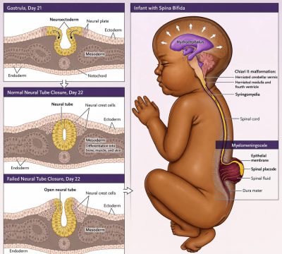
What Causes Myelomeningocele?
The exact cause is unknown. Research suggests that a combination of genetic factors, certain medications, and maternal nutrition may contribute to the condition. Babies may also have a higher risk if the mother has diabetes during pregnancy.
How is Myelomeningocele Diagnosed?
I can often detect myelomeningocele, the most common form of spina bifida, during your routine anatomy ultrasound between 18 and 22 weeks of pregnancy. In some cases, I may also recommend a maternal blood test as early as 16 weeks to measure alpha-fetoprotein (AFP). Elevated AFP levels can be an early sign that your baby may have an open neural tube defect, prompting me to perform a more detailed evaluation.
If I see findings on your ultrasound or your AFP level is higher than expected, I’ll discuss additional tests with you to confirm the diagnosis and better understand your baby’s condition. One of these tests is an amniocentesis, especially if you’re considering fetal surgery. During this procedure, I collect a small sample of the amniotic fluid surrounding your baby and analyze it for markers, including AFP. These results help me confirm whether spina bifida is present, determine the specific type of defect, and guide you through the most appropriate treatment options.
Intrauterine treatment
If your baby is diagnosed with myelomeningocele, I may discuss fetal surgery as a treatment option. Since its introduction in 2011, fetal repair has transformed the care of babies with spina bifida by reducing many of the long-term complications associated with the condition. If you are a suitable candidate, I will carefully evaluate you and your baby, explain the benefits and potential risks, and guide you through every step of the decision-making process so you can make the choice that is right for your family.
Fetal surgery is typically performed between 19 and 26 weeks of pregnancy. During the procedure, a specialized multidisciplinary team carefully opens the uterus to access your baby, repairs the spinal defect, and protects the exposed spinal cord with a specialized waterproof covering to minimize further injury from the amniotic fluid. Once the repair is complete, I will closely monitor both you and your baby throughout your recovery. You’ll usually stay in the hospital for four to five days, followed by weekly clinic visits and ultrasound examinations to ensure that your pregnancy continues safely until delivery.